 |
While specimen preparation of superalloys for metallographic examination is relatively straightforward, the metallographer must take into consideration some of the inherent characteristics of these complex alloys, such as high toughness, presence of large amounts of strengthening phase and high corrosion resistance, to ensure getting samples that clearly reveal their complex microstructures.
Superalloys are complex alloys of Fe-Ni, Ni-, and Co-base compositions. Their microstructure can be quite complex due to the potential for a variety of phases that can form in heat treatment or service exposure conditions. This article discusses the use of new metallographic materials to prepare these alloys and the different etchants required to reveal the structure of these alloys properly as a function of alloy composition, heat treatment and microstructural phases. This discussion is limited to iron-nickel and nickel-base alloys, but most of the comments are also applicable to Co-base alloys.
Preparation of nickel-base superalloys has many similarities to procedures used for stainless steels. Because they are face-centered cubic (FCC) austenitic alloys with exceptional toughness, machinability is poorer than for steels, and age-hardened alloys can be more difficult to section than most steels when they have a high gamma prime (γ′) content. FCC metals readily deform and work harden, so aggressive sectioning methods, such as power-hack sawing or band sawing, can introduce considerable damage, which can be difficult to remove in the subsequent preparation steps. If these procedures must be used, it is advisable to resection the material with the correct abrasive cutoff wheel (consumable type) with abundant cooling. These newly prepared surfaces have less damage and should be used as the starting surface for metallographic preparation.
Mounting
Depending on the nature of the analysis, specimens can be mounted. Mounting is advisable for edge preservation if a surface is to be examined. Compression-mounting thermosetting epoxy resins, such as DuraFast resin, provide the best edge retention. Mounting presses that cool the specimen to ambient under pressure reduce the occurrence and degree of shrinkage gaps that degrade edge retention and lead to bleed-out problems of solvents or etchants and stains, which obscures the edge from examination. Mounting also may be merely a convenience, as the back of the mount is an ideal surface for recording specimen details.
Grinding
Grinding of specimens traditionally has been performed using a series of water-cooled SiC papers of varying grit sizes (e.g., 120, 240, 320, 400 and 600 grit), grinding with each paper for 60-120 seconds at 240-300 rpm on a rotating wheel. The specimen is held against the SiC paper by hand or using a fixture. If done manually, the specimen is rotated 45 to 90 degrees between papers so the grinding direction is not constant. Semiautomatic and automatic grinding machines produce omnidirectional grinding patterns and yield superior flatness and edge preservation. Unless specimens are porous or contain cracks, a simple washing operation between steps is adequate. Otherwise, ultrasonic cleaning with a suitable solvent is recommended.
In contemporary methods, only one grinding step is needed using SiC paper or some other abrasive depending on laboratory throughput or personal preference. In this approach (see Table 1), the best quality metallographic cut-off wheels should be used for sectioning to minimize sectioning damage. Then, use the finest SiC grit size that removes the sectioning damage in a reasonable time. Usually, 180- or 240-grit SiC paper, or products with an equivalent abrasive particle size can be used.
Polishing
In the traditional preparation method, grinding is followed by two to four polishing steps, depending on the quality level needed. Polishing usually commences with either 6- or 3- µm diamond abrasive (paste, slurry, or aerosol) with the appropriate liquid extender/lubricant on a cloth pad. Historically, canvas, billiard, felt, nylon, and synthetic napless or low-nap cloths have been used. Modern processes use napless cloths, such as, Plan, Dur, Dac, and Sat. The choice is often a matter of personal preference or a matter of cost. Wheel speeds are lower for polishing, generally 120-150 rpm. Some operators follow the first polishing step in the traditional method with a second diamond abrasive step (generally 1 µm), or they use one or two steps with aqueous aluminum-oxide slurries. Cloths and wheel speeds are similar, and each step is 60-120 seconds. Cleaning between polishing steps must be performed carefully.
The most common alumina abrasive sizes are 0.3 µm (alpha alumina) and 0.05 µm (gamma alumina). Recently, acidic alumina slurries and basic colloidal silica slurries have been used for final polishing of superalloys, which are highly effective. Age hardened superalloys are easily polished damage-free with standard grinder/polishers. If it is in the solution annealed condition, following preparation with a brief vibratory polish may be helpful. Final polishing cloths may be synthetic and napless, such as OP-Chem, or may have a short nap, such as Floc, Nap or Plus.
Contemporary practices, use flat, napless cloths and pads, except for the final step. But synthetic, napless polyurethane pads can be used even in the final step. These procedures rely on use of automated devices to improve specimen flatness and reproducibility of the procedure.
 |
| Table 1 |
Table 1 shows an example of a contemporary procedure for Ni-base superalloys. The five-step method produces excellent surfaces to observe the true structure. It is required for examining solution-annealed grades, but a four-step procedure can be used with age-hardened grades (drop the 1-µm step). Stretching cloths over a platen, as done in the Traditional Method, cannot be used with semi-automated or fully automated systems as the head will rip a stretched cloth off. Instead, use either psa (pressure-sensitive adhesive) cloths attached to a ferromagnetic disc, or cloths permanently bonded to a steel disc, which are then placed upon the magnetic material on the platen. Charge cloths (especially when new) with diamond in paste form, add Green or Red lubricant and start polishing. If a new cloth is charged with diamond in slurry form, the cutting rate will be low initially until the particles become embedded in the cloth surface. For final polishing, it is best to use an alumina slurry, such as OP-AN, rather than colloidal silica, if a Cl-containing etching is to be employed (etching problems will result). If colloidal silica is used for the final polish, follow with a brief polish with alumina.
Etching
 |
|
Fig 7 Grain structure of solution-annealed Alloy 625 bar (191 HV); acetic glyceregia etch; magnification bar is 100 µm long
|
 |
| Table 2 |
Numerous etchants have been used to reveal the structure of superalloys. However, as for most metals, it is wise to examine the polished surface carefully before etching. Some minor second-phase particles can be easily observed in the as-polished condition using bright field illumination, and nonmetallic inclusions can be best observed in the as-polished condition. Most wrought superalloys are double vacuum-melted (VIM/VAR) and have extremely low sulfur and oxygen contents and, thus, very low inclusion content. The sulfur content in many alloys is well below 0.001 wt%, and no sulfides are observed. Cast superalloys are generally made by vacuum induction melting (VIM) and contain higher inclusion content (and more nitrides and carbides) than wrought alloys.
Several etchants commonly used to reveal the general structure of superalloys are listed in Table 2. Due to the excellent corrosion resistance of superalloys, most etchants work best by swabbing (use a light pressure) the specimen with cotton soaked in the etchant. Immersion etching often results in a more irregular etch response. Many of the etchants must be mixed fresh and used within a short time span. Glyceregia is the mildest of the first three etchants in Table 2, and etchant 2 or 3 can be used if etching occurs too slowly. Glyceregia is good for revealing second-phase precipitates but not particularly good for developing the grain and twin boundaries.
Microstructure
Fe-Ni and Ni-base superalloys have an austenitic (γ) phase matrix and do not exhibit allotropy. They are designed for use above ~540°C (1000°F), but often are used in low-temperature applications due to their exceptional toughness. Numerous second-phase particles can be observed; for example, a variety of carbides (MC, M23C6, M6C and M7C3 types), nitride and strengthening precipitates, γ′ or γ″. In general, γ′ and γ″ are too small to see with the light microscope unless the specimen is over-aged substantially. Gamma prime (γ′) is found in nickel-base alloys strengthened by addition of Al and Ti. It is an ordered face-centered cubic precipitate of the form Ni3(Al,Ti). Gamma double prime (γ″) is found in nickel-base alloys strengthened by additions of Nb (Cb), such as Alloy 718. It is a body-centered tetragonal phase of the form Ni3Nb. Excessive high-temperature exposure can produce other phases such as the hexagonal close-packed eta phase (Ni3Ti) and the orthorhombic delta phase (Ni3Nb). Laves phase (Fe2Nb, Fe2Ti or Fe2Mo) may be encountered in some alloys, particularly in the as-cast and as-rolled conditions.
 |
|
Fig 9 Partially recrystallized Waspaloy grain structure; 1010°C solution anneal and double aged; modified Beraha’s tint etch; mag. bar is 100 µm long
|
Because these alloys are not allotropic, control of grain size is more difficult than for heat treatable steels. The hot-working temperatures must be carefully controlled to yield a fine-grained structure. As an example, Fig. 1 shows the as-rolled microstructure of Custom Age 625 Plus [1] finish rolled at 1007°C (1845°F), which yielded a very fine grain structure. Solution annealing of this material at 968°C (1775°F) resulted in only a small degree of recrystallization and grain growth (Fig. 2).
Solution annealing at 1010°C (1850°F) produces a fully recrystallized structure with some grain growth (Fig. 3).
However, control of grain size in hot working is difficult, and nonstandard structures do occur. For example, Fig. 4 shows a partially recrystallized duplex grain structure in an as-forged specimen of a 625-type alloy composition. This condition is referred to as a “necklace” type duplex condition, where large nonrecrystallized grains are surrounded by fine recrystallized grains. This is one of several types of duplex grain size conditions that are encountered in superalloys. Large as-forged specimens can also exhibit rather complete (or less complete) grain boundary carbide networks as illustrated in Fig. 5. This structure is from the center of a 305-mm (12-inch) diameter as-forged bar of Alloy 600.
Solution annealing dissolves these grain boundary carbide films, which usually consist of carbide types (M23C6 carbide, for example) that are more easily dissolved. However, other carbide types (MC-type carbides, for example) resist solutioning up to the melting point. Figure 6 shows the fine MC precipitates in a 140-mm (5.5-inch) diameter bar of Alloy 625 after solution annealing at 982°C (1800°F). The grain structure of this specimen is shown in Fig. 7.
High temperature exposure can result in precipitation of undesirable phases. Figure 8a shows precipitation of (η) eta phase (Ni3Ti) in Pyromet 31, an Fe-Ni-base superalloy, after exposure at 816°C (1500°F) for 1,500 hours. Figure 8b shows eta phase in another Pyromet 31 specimen solution annealed at 954°C (1750°F). It is much finer but has not been completely put into solution at this temperature.
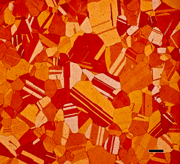
Control of the solution annealing temperature is important. A low solution annealing temperature keeps the grain structure fine (although it may not be fully recrystallized), but may not dissolve undesired phases such as grain boundary carbides, eta and delta. On the other hand, a high solution annealing temperature dissolves these phases and recrystallizes the grain structure, but will permit substantial grain growth. Choice of solution annealing temperature also is governed by the desired end properties; that is, whether toughness, stress rupture, or creep properties are most important. Figures 9 – 11 demonstrate how the solution annealing temperature influences recrystallization and grain growth. These are solution-annealed microstructures of Waspaloy, all given a standard double aging treatment. The specimens were tint etched in a modified Beraha’s solution (immersed in 50 ml HCl, 50 ml water, 1 g potassium metabisulfite and 2 g ammonium bifluoride until the surface was colored violet). Color etching of Ni-based superalloys is very difficult.
Figure 9 shows the grain structure after a 1010°C (1850°F) solution anneal, which is only partially recrystallized. Figure 10 shows the grain structure after a 1038°C (1900°F) solution anneal, which is fully recrystallized and somewhat coarse. Figure 11 shows that solution annealing at 1066°C (1950°F) produces only a small degree of additional grain growth. Note the excellent development of the grain structure using a tint etch. In comparison, the specimen solution annealed at 1038°C (Fig. 10) is not as well developed by etching with glyceregia as shown in Fig. 12. This was obtained after swabbing for several minutes.
Alloy 718 is the most popular Ni-base superalloy. Various aspects of its heat treatment and microstructure are illustrated in Figs. 13 and 14. Figure 13 shows precipitates present after solution annealing at a low temperature of 954°C (1750°F), which yielded a hardness of 89 HRB, but delta phase (δ) is present along the grain boundaries. The globular particles are MC carbides. Figure 14 shows the grain structure after solution annealing at 1066 C. Note that the grain and twin boundaries are free of delta phase. Only carbides and nitrides are visible. The hardness is 80 HRB. Aging this specimen increases hardness. A double aging treatment usually is used; the first aging treatment is an 8 hour hold at 718°C (1325°F), which increases the hardness to 29 HRC. After this age, the specimen is cooled at a slow rate to 621°C (1150°F), held for an additional 8 hours, then air cooled to ambient temperature, which increased the hardness to 37 HRC. However, no visible change can be observed in the light micrographs compared with the solution annealed condition (Fig. 14). The γ″ strengthening is best observed with a TEM using dark field.
The γ′ or γ″ strengthening phase in superalloys heat treated in the prescribed manner is always too fine to see with the light microscope. In some systems, it is possible to see γ′ with an SEM even without a field emission gun. The γ″ in some alloys (Custom Age 625 Plus, for example), is much too fine to be observed with the SEM unless the specimen has been over-aged. During metal processing, there are times when γ′ is coarse enough to be observed with the light microscope. Such an example is given in Fig. 15, which shows γ′ (and grain boundary carbides) in as-HIP’ed (hot-isostatically pressed) René 95, a powder metallurgy processed grade.
George Vander Voort has a background in physical, process and mechanical metallurgy and has been performing metallographic studies for 43 years. He is a long-time member of ASTM Committee E-4 on metallography and has published extensively in metallography and failure analysis. He regularly teaches MEI courses for ASM International and is now doing webinars. He is a consultant for Struers Inc. and will be teaching courses soon for them. He can be reached at 1-847-623-7648, georgevandervoort@yahoo.com and through his web site: www.georgevandervoort.com
To View a listing of all George’s articles please click here
Read George Vander Voort‘s Biography


