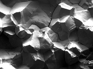
Commercial introduction of the Scanning Electron Microscope (SEM) in 1965, and its subsequent rapid development and implementation in metallographic laboratories, has had a profound influence on failure studies. The chief advantage of the SEM is its great depth of field in comparison of the light microscope. Observations can be made over a much wider range of magnifications including those above the range for light microscopes (and those below the range of TEM replicas). Examination of fracture features by SEM is much simpler than through the study of replicas with the Transmission Electron Microscope, TEM. A further advantage of the SEM is the chemical analytical capability of spectrometers that can be attached to the microscope, Energy-dispersive Spectrometers (EDS) being the most common.
The study of fractures with the unaided eye has been practiced since antiquity chiefly for controlling the quality of metals production. R. A. F. de Réammur may have been the first to examine and publish drawings of fractures examined at high magnifications, at least 100X, in 1722. R. Mallet appears to have been the first to link fracture appearance to service performances in a study of failed cannon barrels published in 1856. Adolf Martens may have been the first to study both fracture surfaces and the underlying microstructures in 1878 followed by the first description of fracture surface features in 1887 when he showed that these lineal features could be traced backward to identify the fracture origin.



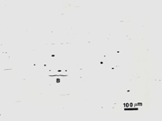 Over the years, ASTM Committee E-4 on Metallography has conducted interlaboratory test programs to evaluate the precision and bias associated with measurements of microstructure using proposed and existing test methods. ASTM decided in the late 1970s that all test methods that generated numerical data must have a precision and bias section defining the repeatability and reproducibility of the method. Defining bias associated with a test method is difficult unless there is an absolute known value for the quantity being measured and this is not possible when microstructural features are being measured. This paper shows the results for an interlaboratory test using Method A, “worst field” ratings of inclusions in steels by ASTM E-45. The results from 9 people who were reported to be qualified, regular users of the method revealed consistent problems of misclassification of inclusions types and a wide range of severity ratings for each specimen.